您當前的位置:檢測資訊 > 科研開發
嘉峪檢測網 2022-07-08 11:36
理想的骨修復材料除具備良好的生物學活性與力學性能外,還應具備內部連通的多孔結構與個性化的幾何外觀。本研究采用選擇性激光熔化技術制備了力學性能適配的3D打印多孔Mg-Nd-Zn-Zr骨修復支架,并通過體內外實驗研究其生物安全性與抗菌性能。
01、研究內容簡介
金屬植入物被廣泛應用于骨科領域。植入物感染是骨科手術后最嚴重的并發癥之一。傳統上,植入物感染的治療方案包括手術清創、全身抗生素治療和局部抗生素治療等,將不可避免地導致二次手術及各類并發癥,也造成了嚴重的社會經濟負擔。因此,亟需研發一種具備抗感染性能的骨植入物材料用于預防和治療此類感染。
鑒于其良好的生物相容性、骨誘導性和力學性能,可降解鎂合金被視為新一代骨修復材料。既往研究表明,鎂合金降解產生的堿性微環境具備廣譜殺菌性能,可有效抑制革蘭氏陽性及革蘭氏陰性細菌的生長。因此,鎂合金作為一種兼具抗菌和成骨性能的可生物降解材料,具有良好的臨床轉化前景。
理想的骨修復材料應具備內部連通的多孔結構與個性化的幾何外觀。目前,多孔鎂合金植入物的主要制備方法包括熔模鑄造法、粉末冶金法、溶體發泡法等方法,能夠制備具有完全內部連通多孔結構的鎂合金骨修復支架,但無法實現多孔鎂合金內部孔隙結構的精準控制。近年來,金屬增材制造技術的發展,為制備孔隙結構可調、內連通性可控的鎂合金骨修復支架提供了新思路。然而,鎂金屬化學性質活潑,極易燃,采用激光或電子束制備3D打印鎂合金存在諸多挑戰,其體內生物學作用與功能亦未見報道。因此,我們團隊通過醫工交叉合作,采用選擇性激光熔化技術,制備3D打印多孔Mg-Nd-Zn-Zr骨修復支架,并通過體內外實驗系統評估其抗菌性能與生物安全性,為3D打印鎂合金植入物的研發與臨床轉化提供依據。
設計具有鉆石型結構單元的多孔骨修復支架,支架設計孔徑為400μm。支架設計完成后,導出slt格式打印文件,用于后續打印。通過氣體霧化法成功制備了Mg-Nd-Zn-Zr鎂合金粉末,粉末球型度高,平均粒徑為63.9 ± 14.5μm,符合3D打印要求(Fig 1)。
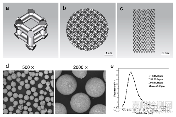
Fig. 1. Design of the 3D-printed JDBM implant and characterization of Mg–Nd–Zn–Zr powder. a) Diamond unit cell. b) Top view of the computer-aided design model. c) Lateral view of the computer-aided design model. d) SEM image of the Mg–Nd–Zn–Zr powder. e) Particle size distribution of the Mg–Nd–Zn–Zr powder.
經后處理后,3D打印多孔Mg-Nd-Zn-Zr支架多孔結構分布均勻,324.6±25.7μm,孔隙率為 52.1±1.6%。同時,通過體外浸泡實驗,初步評價3D打印多孔Mg-Nd-Zn-Zr支架的降解性能(Fig 2)。
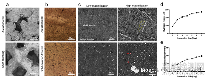
Fig. 2. Characterization and degradation behavior of the 3D-printed JDBM implant. a) Surface morphology of the 3D-printed JDBM implants. b) Optical microscope images of 3D-printed JDBM implants. c) SEM images of as-fabricated and solid-solution-treated 3D-printed JDBM implant observed from side surfaces. d) Hydrogen release after immersing for 1–7 days. e) Mg2+ concentrations after immersing for 1–7 days.
體外實驗結果表明,3D打印多孔Mg-Nd-Zn-Zr支架具備良好的生物安全性與促成骨活性(Fig 3)。
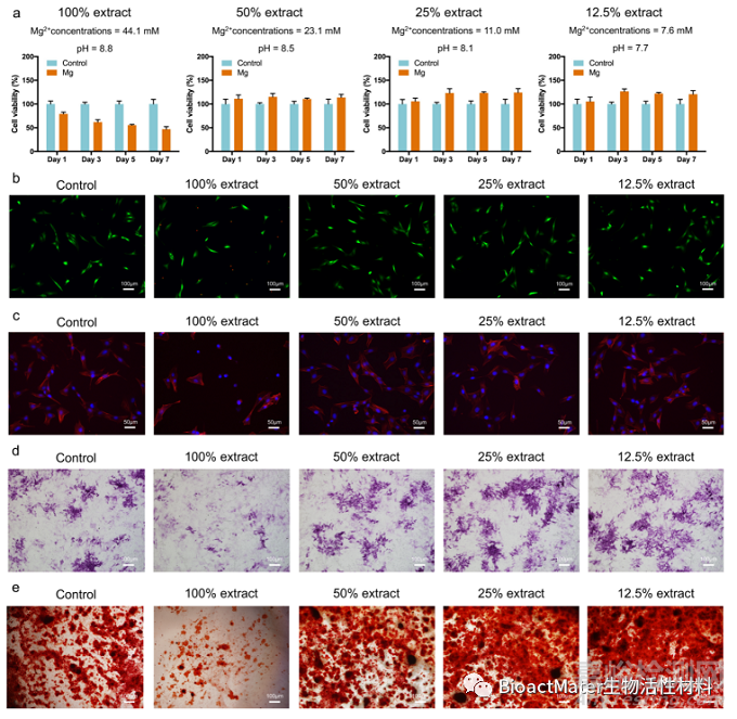
Fig. 3. Cytocompatibility and osteogenic differentiation in vitro. a) Cell viability, b) live/dead cells, and c) cell morphology of the MC3T3-E1 cells cultured with different extract concentrations. d) ALP staining after 7 d of culture. e) Alizarin red staining of cells cultured with different extract concentrations for 21 d.
3D打印多孔Mg-Nd-Zn-Zr支架可顯著抑制MRSA 粘附及細菌生物膜形成(Fig 4)。

Fig. 4. Antibacterial activity of 3D-printed JDBM implants in vitro. a) Biofilm formation of MRSA cultured in different media after 1, 3, 6, 12, and 24 h of incubation. b) Absorption of crystal violet by MRSA biofilm after 24 h of incubation. c) Semi-quantitative analysis of bacterial concentration after 1, 3, 6, 12, and 24 h of in- cubation. d) Representative images of MRSA cultured in different media. e) Statistical analysis of colony number after 24 h of incubation. *p < 0.05.
動物實驗結果表明,3D打印多孔Mg-Nd-Zn-Zr支架可有效預防植入物感染形成(Fig 5)。
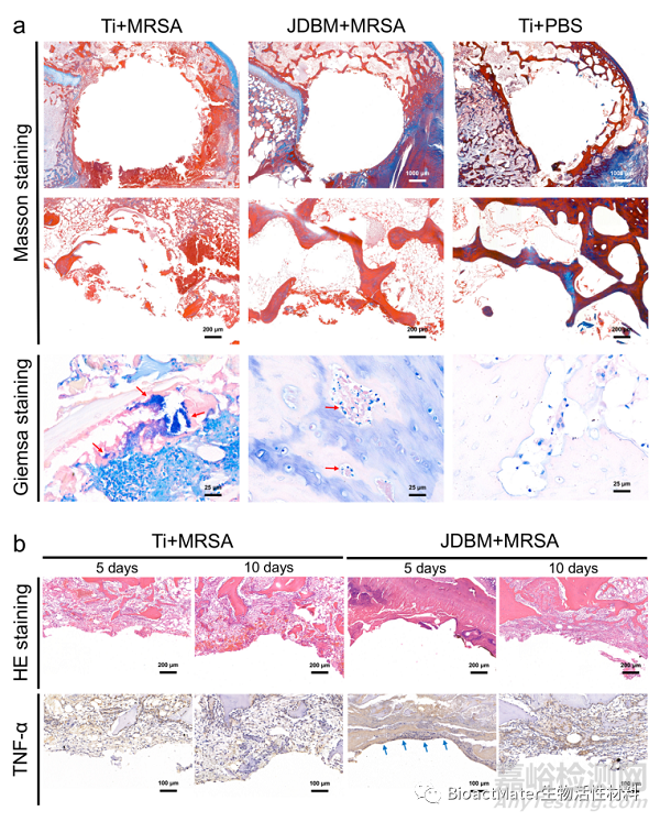
Fig. 5. Histological evaluation of implant-related infection. a) Masson and Giemsa staining of peri-implant tissue at four weeks after implantation. b) HE staining and immunohistochemical detection of TNF-α secretion at the bone-implant interface at 5 and 10 days after implantation.
此外,3D打印多孔Mg-Nd-Zn-Zr支架具備良好的生物安全性。3D打印多孔Mg-Nd-Zn-Zr支架植入后,實驗動物心、肝、脾、肺、腎未見 明顯組織學異常改變(Fig 6),實驗動物主要臟器鎂離子含量未見異常升高(Table 1)。
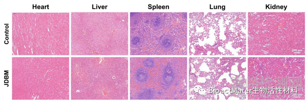
Fig. 6. Evaluation of organizational microstructures of the heart, liver, spleen, lung, and kidney with HE staining.
Table 1 Results of blood tests of experimental animal at 4 weeks after implantation.
|
Cr |
ALT |
AST |
ALB |
A/G |
|
|
Control |
69.2 9.2 |
39.8 .4 |
21.1 |
49.3 |
2.2 |
|
JDBM |
76.6 9.4 |
37.4 9.7 |
21.2 4.5 |
47.4 |
2.5 |
|
p value |
0.363 |
0.771 |
0.972 |
0.652 |
0.710 |
|
Cr |
ALT |
AST |
ALB |
A/G |
|
|
Control |
69.2 9.2 |
39.8 .4 |
21.1 |
49.3 |
2.2 |
|
JDBM |
76.6 9.4 |
37.4 9.7 |
21.2 4.5 |
47.4 |
2.5 |
|
p value |
0.363 |
0.771 |
0.972 |
0.652 |
0.710 |
綜上所述,在這項研究中,我們通過選擇性激光熔化技術實現了3D打印多孔Mg-Nd-Zn-Zr支架的制備,通過體內、 外實驗證實其具備良好的生物安全性與抗感染性能,為其臨床轉化提供依據。

來源:BioactMater生物活性材料


